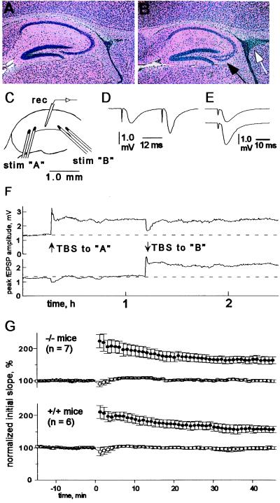
Figure 3.
Hippocampal morphology and physiology. (A and B) Morphology of adult hippocampus. Hematoxylin and eosin stain (×14) of wild-type (A) and homozygous (B) adult brain. Hippocampal physiology: (C) Typical stimulus (stim) and recording (rec) electrode positions. (D) Example of fEPSPs elicited with paired stimulation; interpulse interval equals 25 ms. Waveforms (E) are sample fEPSPs collected immediately before (upper trace) and 40 min after (lower trace) TBS. (F) Typical record of LTP induced in slices from knockout mice. The upper and lower traces are amplitudes of fEPSPs elicited alternately at 0.1 Hz along two independent pathways in the stratum radiatum of field CA1. Delivery of TBS to path A (orthodromic) resulted in a rapid enhancement of fEPSP amplitudes that decayed over a brief period to a stable plateau. Approximately 1 h later, application of the same high-frequency stimulus to pathway B (antidromic) resulted in a comparable enhancement, without affecting established LTP in path A. The initial period of enhancement after TBS is thought to reflect a mixture of post-tetanic potentiation and short-term potentiation, a more decremental form of N-methyl-d-aspartate (NMDA) receptor-dependent plasticity. The brief heterosynaptic depression observed after TBS is likely related to adenosine release accompanying high-frequency stimulation. (G) A plot of the average amount of LTP obtained in groups of slices from N-CAM knockout and wild-type mice. TBS-induced potentiation, expressed as a percentage of the preconditioning EPSP slope, was similar in the two groups of slices with regard to time course, magnitude, and input specificity (n = number of animals). Amplitudes of response to both the conditioned (•) and nonconditioned (□) pathways are shown.

