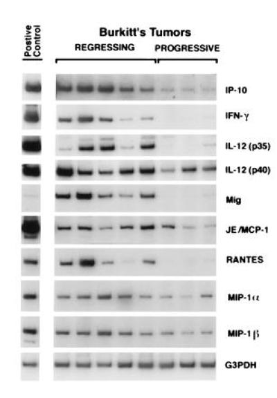
Figure 1.

Cytokine and chemokine mRNAs in regressing and progressive Burkitt tumors revealed by RT-PCR analysis. Tissue fragments were obtained from regressing and progressive Burkitt tumors established in athymic mice. Positive controls were derived from murine macrophages incubated with 1 mg/ml lipopolysaccharide for 6 hr (IP-10, IL-12, Mig, and JE/MCP-1); murine splenocytes incubated with 4 mg/ml concanavalin A for 3 hr (IFN-γ); murine splenocytes from a 3-day mixed lymphocyte reaction (RANTES, MIP-1α, and MIP-1β). Total cellular RNA, isolated from tumor and control tissues, was subjected to RT-PCR analysis. After normalization to G3PDH, the signals derived from cytokine expression in regressing versus progressive Burkitt tumors, expressed as ratios, were as follows: IP-10, 8.2; IFN-γ, 10.6; IL-12 p35, 15.6; IL-12 p40, 4.0; Mig, 12.8; JE/MCP-1, 2.3; RANTES, 8.1; MIP-1α, 2.8; MIP-1β, 2.7.
