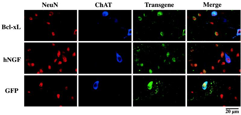
Figure 2.
Immnohistochemical staining of coronal sections through the septal area. Confocal microscopic images show in vivo transduction of adult rat septal neurons expressing neuronal markers (NeuN; red), cholinergic markers (ChAT; blue), and the hNGF or Bcl-xL transgene (hNGF/Bcl-xL; green). The images obtained from each individual staining and from the merged images are shown. Representative fields of the area surrounding the injections site are shown with several cells triple labeled for hNGF, NeuN, and ChAT, as well as for Bcl-xL, NeuN, and ChAT.

