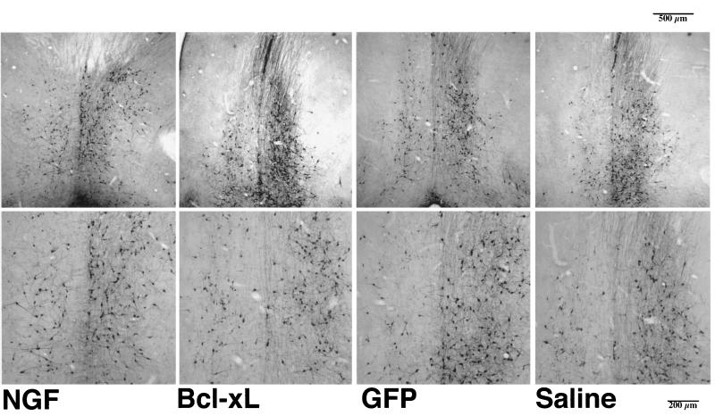
Figure 3.
Coronal sections (50 μm) through the medial septum stained for ChAT immunoreactivity 6 weeks after hNGF, Bcl-xL, or GFP vector injection or saline control injection and 3 weeks after unilateral aspirative lesion of the fimbria fornix, ipsilateral to the injection. The medial septum was defined as the area lying above a line between the midportion of the anterior commissure, beneath the corpus callosum, and laterally limited by the ventricles. ChAT-positive cells displaying a round cell body and at least one process were counted for the lesioned and the unlesioned site of the septum.

