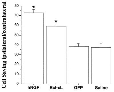
Figure 4.
Percentage of ipsilateral/contralateral septal neurons quantified after intraseptal vector injections and lesion of the fimbria fornix. The percentage of ChAT-immunoreactive cells (mean ± SEM) are shown for hNGF-, Bcl-xL-, and GFP-expressing vector and saline control. hNGF and Bcl-xL viral vector-injected animals presented significantly higher numbers of surviving cholinergic septal neurons compared with GFP viral vector or saline-injected control animals (∗, P < 0.05, ANOVA, Fischer post hoc test).

