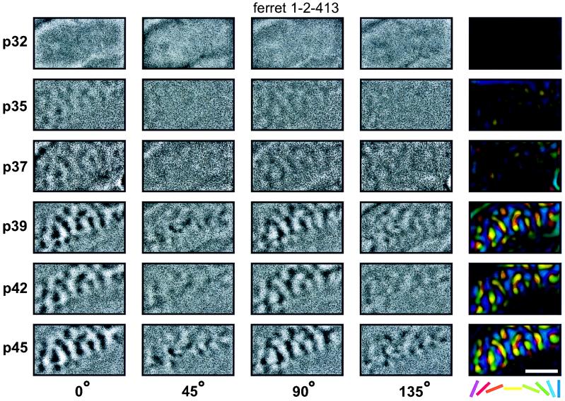
Figure 1.
Faster development of horizontal and vertical than of oblique orientation maps. Each row of the figure shows orientation maps recorded in the left primary visual cortex of one ferret at the age (postnatal day, p) indicated at the left of the row. The first four columns show orientation maps recorded in response to a particular orientation of moving square-wave grating (0° = horizontal). The fifth column shows the polar map calculated from those four single-condition maps. For each map, caudal is up and medial is to the left. The curve in the upper left corner of each map indicates the location of the caudal pole of the cortex behind which the skull remained intact over the cerebellum. The approximate location of the area 17/18 border can be seen in each image as a line rostral to which orientation activity is not seen. Single-unit recordings were not performed in this study to precisely locate the area of the visual field studied in each animal. However, the craniotomies were always performed at the same location on the skull with respect to skull sutures, exposing what has previously been found to be the representation of area centralis (2, 3). In this example the first orientation maps can be seen at postnatal day 35. At this age only horizontal and vertical maps are clearly seen. The orientation maps for the two oblique orientations develop considerably slower (they are first clearly seen at p37) and the difference in the strength of the maps remains present throughout the experiment. (Bar = 2 mm.)

