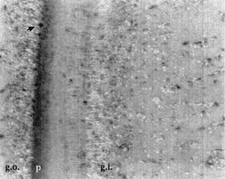
Figure 4.

Immunohistochemical localization of CE in the squid optic lobe. Sections were incubated with CE antibody and visualized by immunoperoxidase staining. Nuclei were counterstained blue with hematoxylin (g.o., outer granule cell layer; g.i., inner granule cell layer; p, plexiform layer). Arrow points to an amacrine cell. The plexiform layer containing neuronal axons and branches is darkly stained with the immunolabel.
