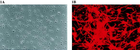
Figure 1.
Morphology and characterization of cultured feline astrocytes. (A) Phase–contrast micrograph of primary astrocyte cultures maintained with DMEM supplemented with 10% fetal bovine serum for 6 weeks (×100). (B) Cultured astrocytes stained with rabbit anti-bovine GFAP and visualized by immunofluorescence with donkey anti-rabbit lissamine rhodamine B sulfonyl chloride-conjugated (×200).

