Figure 4.
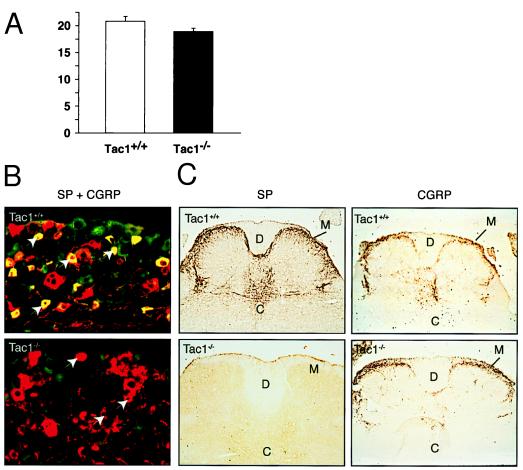
Substance P producing cells in DRGs and spinal cord. (A) Neurons were marked using a specific neuronal nuclear marker (NeuN) [chemicon (1:2000)], and the number of positive nuclei in several sections of 4–6 DRGs per mouse were counted using one square inch grid. The number of neurons in the DRGs from Tac1+/+ (n = 3) and Tac1−/− (n = 3) mice. (B) Double staining of DRGs from Tac1+/+ and Tac1−/− mice with substance P and CGRP specific immunosera. Substance P-positive cells typically have small diameters and are labeled green (fluorescein isothiocyanate), while CGRP positive cells are labeled red (CY3). Double-positive cells therefore appear yellow. Arrows point to small-diameter CGRP-positive and substance P-negative cells in the DRG of Tac1−/− mice. (C) Analysis of substance P and CGRP immunostaining in the spinal cord. The presence of both peptides, mainly in lamina I and II, overlaps in wild-type Tac1+/+ mice. Note the absence of substance P immunoreactivity in Tac1−/− animals, while the CGRP-staining is normal. C, central canal; D, dorsal columns; M, marginal zone of the dorsal horn.

