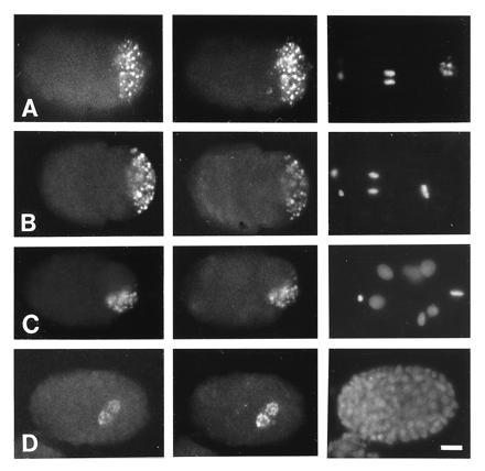
Figure 4.

Immunofluorescence staining of embryos with OIC1D4 and anti-GLH-1 and anti-GLH-2 antibodies. Embryos are oriented with anterior left and ventral down. (A). Embryo stained with affinity-purified chicken anti-GLH-1 antibodies (Left), mouse monoclonal antibody OIC1D4 (Center), and DAPI (Right). (B–D). Embryos stained with affinity-purified mouse anti-GLH-1 (Left), chicken anti-GLH-2 (Center), and DAPI (Right). (A and B) Two-cell embryos. P1 is in mitosis, and P granules are segregated to the posterior cortex destined for P2. (C) Seven-cell embryo. P2 is in mitosis, and P granules are segregated to the ventral region destined for P3. (D) Late-stage embryo showing P granules in Z2 and Z3. (Bar = 10 μm.)
