
Figure 2.
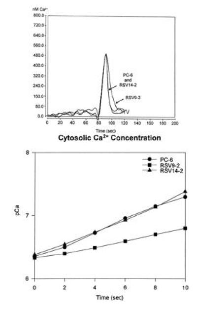
Bradykinin-induced calcium flux measurements in wild-type and transfected PC6 cells. Cells were scrape-loaded with aequorin and allowed to attach overnight to the surface of the Sykes–Moore chambers. The cells were then placed in a luminometer and changed into calcium- and serum-free medium; bradykinin (100 nM) was applied and the luminescence was monitored. (Upper) The calcium transients measured for each cell line. (Lower) A semilog plot of the rate of Ca2+ removal for each of the cell lines between 90 and 100 sec).