Figure 2.
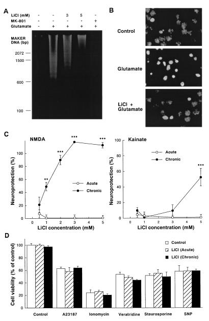
Effects of chronic lithium pretreatment on neurotoxicity of cerebellar granule cells. (A) Chronic lithium prevents glutamate-induced internucleosomal DNA fragmentation of cerebellar granule cell. Cells were pretreated with various concentrations of LiCl (3–5 mM) for 7 days and then exposed to glutamate (100 μM) as described in Fig. 1. Soluble DNA was extracted from cells 24 h after the addition of glutamate, subjected to agarose gel electrophoresis, stained with ethidium bromide, and photographed. (B) Chronic lithium inhibits glutamate-induced chromatin condensation of cerebellar granule cell. Cells were pretreated with LiCl (3 mM) for 7 days and then exposed to glutamate (100 μM). Chromatin condensation was detected by nucleus staining with Hoechst 33258. Nuclei were photographed with a Zeiss Axiophot fluorescence microscope at a magnification of ×1,000. (C) Chronic lithium pretreatment protects cells against NMDA and kainate toxicity. Lithium chloride (0.5–5 mM) was added to cultured neurons 1 h (acute) or 7 days (chronic) before NMDA (1 mM) or kainate (100 μM). Additionally MK-801 (1 μM) was added to the kainate-treated groups. After 24 h of stimulation, neuronal viability was measured with the MTT colorimetric assay. The results are expressed as percent of neuroprotection. (D) Effects of lithium on A23187, ionomycin, veratridine, staurosporine, and sodium nitroprusside (SNP)-induced neurotoxicity. Lithium chloride (3 mM) was added to cultured cerebellar granule neurons 1 h (acute) or 7 days (chronic) prior to their exposure to A23187 (300 nM), ionomycin (3 μM), veratridine (1 μM), staurosporine (30 nM), and SNP (100 μM). Neuronal survival was determined 24 h after addition by using the MTT colorimetric assay. Data are the mean ± SEM of viability measurements from four or five cultures. ∗∗, P < 0.01; ∗∗∗, P < 0.001, compared with the group of control (one-way ANOVA with Bonferroni–Dunn test).

