
FIGURE 2.
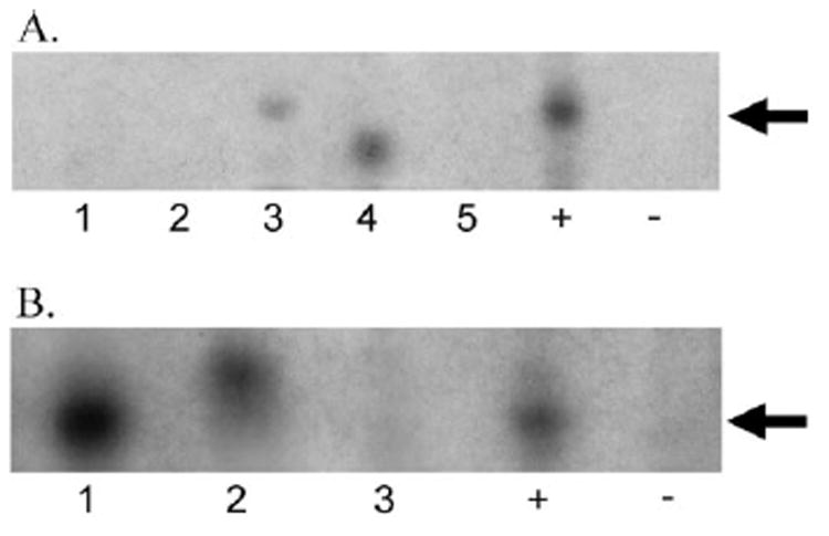
(A) Sample gel of PCR analysis for the bcl-2 t(14;18) translocation at the Mbr. PCR amplification of DNA from microdissected cells from vitreous specimens of PIOL patients using primers for the bcl-2 t(14;18) translocation at the Mbr. Lanes 1 to 5, PIOL samples; +, positive control; −, negative control. Seventy-two vitreous specimens were examined; 40 (55%) were positive. (B) Sample gel of PCR analysis for the bcl-2 t(14;18) translocation at the mcr. PCR amplification of DNA from microdissected cells from vitreous specimens of PIOL patients using primers for the bcl-2 t(14;18) translocation at the mcr. Lanes 1 to 3, PIOL samples; +, positive control; −, negative control. Sixty-eight vitreous specimens were examined; 15 (22%) were positive. In both (A) and (B), differences in migration through the gel were caused by variability where the translocation could occur in the JH region of the IgH gene, resulting in different sizes of amplified product.