
Figure 5.
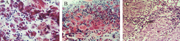
LCNK SCC, A- Fibres cells (arrow) and koilocytes (arrow head), Pap stain, × 400. B- Marked nuclear pleomorphism and hyperchromasia and heavy neutrophilic infiltrate, Pap stain, × 400. C- Corresponding biopsy: islands of neoplastic cells surrounded by heavy neutrophilic infiltrate, H&E, ×10