
Figure 3.
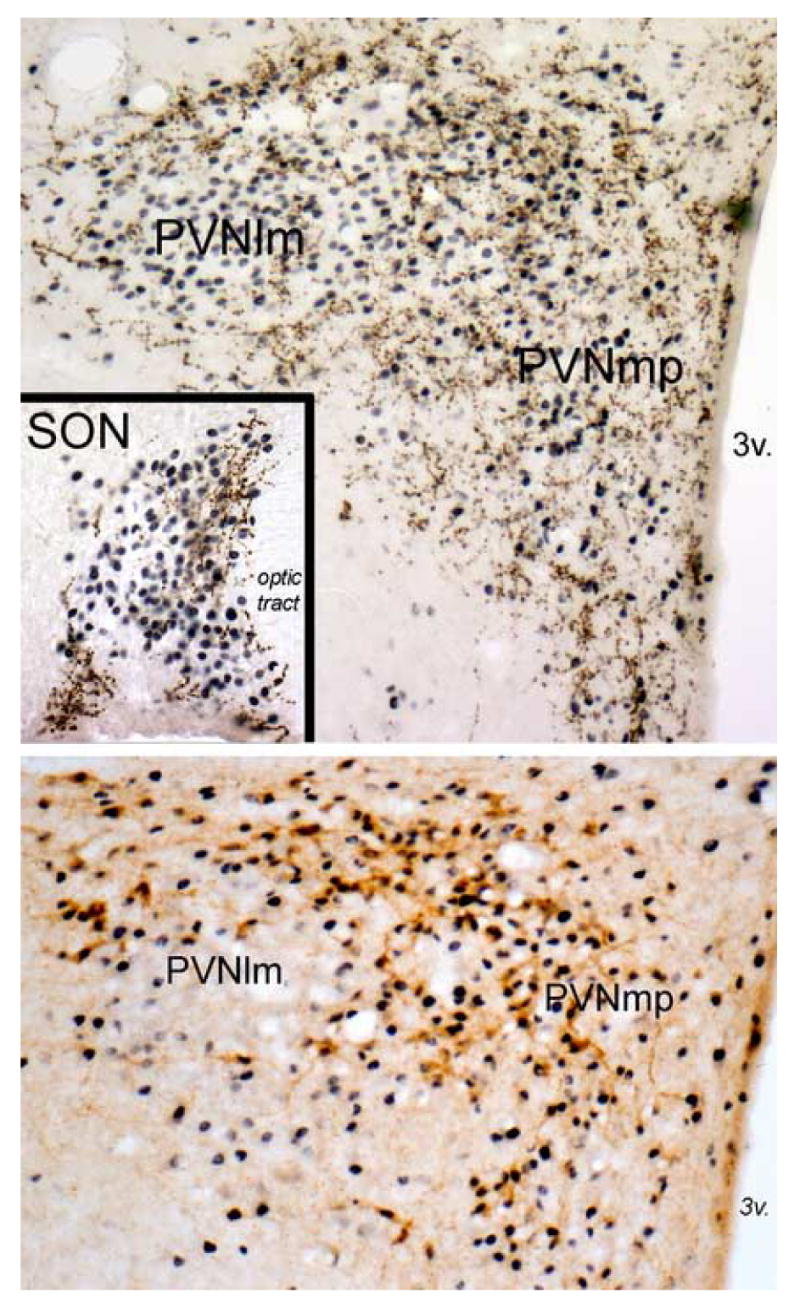
The top panel and inset depict immunoperoxidase labeling of GLP-1 immunopositive fibers (brown) within the PVN and SON (inset) in an adult rat after systemic administration of LiCl (0.15M, 2% BW, i.p.). Robust LiCl-induced neural Fos expression (blue-black nuclear label) is present throughout the PVN, including the PVNmp and PVNlm. GLP-1-positive fibers are largely absent within the core of the PVNlm, where AVP-positive neurons cluster in rats. Instead, GLP-1-positive fibers are distributed around the perimeter of the PVNlm, where magnocellular OT neurons predominate, and throughout the PVNmp, where parvocellular CRF and OT neurons predominate. LiCl-induced Fos expression also is prevalent throughout the SON (inset), where GLP-1-positive fibers cluster within the dorsal and medial SON where magnocellular OT neurons predominate. The lower panel depicts immunoperoxidase labeling of CRF-positive neurons (brown) within the PVN in an adult rat perfused 90 min after intracerebroventricular infusion of 1.0 μg of synthetic GLP-1-(7–36) amide. Fos expression is robust within the PVNmp, including activation of the majority of CRF-positive neurons. Conversely, Fos is largely absent within the core of the PVNlm, where magnocellular AVP neurons predominate. 3v, third ventricle. See abbreviation list.