
Fig. 2.
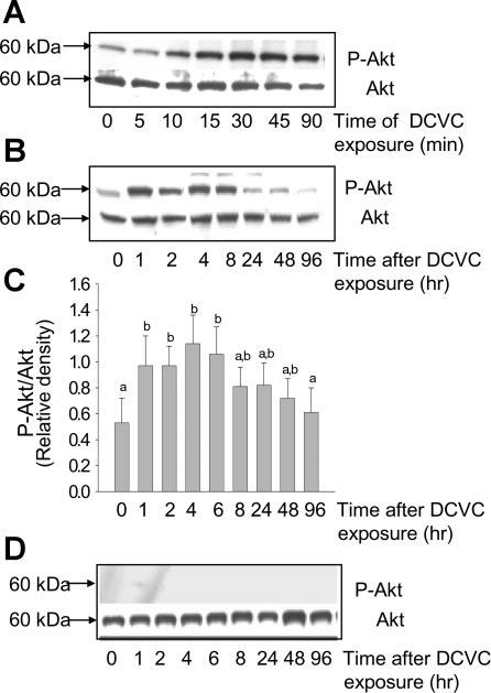
Phosphorylation of Akt(Ser473) in DCVC injured RPTC. A: protein levels of phosphorylated and total Akt in RPTC at different time points of DCVC (240 μM) exposure (90 min). B: protein levels of phosphorylated and total Akt in RPTC at different time points after DCVC exposure. C: ratio of phospho-Akt(Ser473) to total Akt at different time points following DCVC injury in RPTC. The results (quantified by densitometry) are means ± SE (n = 3). Values with dissimilar superscripts (a, b) are significantly (P < 0.05) different from each other. D: effect of PI3 kinase inhibitor (LY294002, 20 μM) on Akt phosphorylation following DCVC injury in RPTC. Blots are representative of 4 independent experiments.