
Figure 3. Levels of expression of ALK in different human breast cancers.
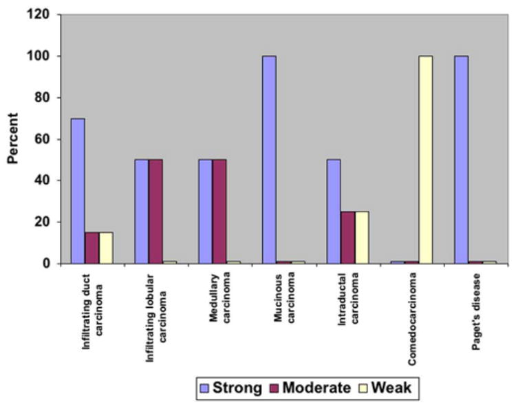
The levels of expression of ALK were quantified using light microscopy and scored in a scale from 1 to 3. The results demonstrated that 75% infiltrating duct carcinomas, 50% infiltrating lobular carcinomas, 50% medullary carcinomas, 100% mucinous carcinomas, 50% intraductal carcinomas, and 100% of the cases of Paget’s disease express high levels of ALK.