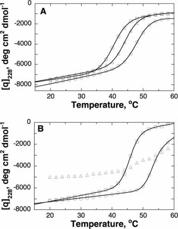
Fig. 5.

Thermal unfolding of wild-type and mutant TyrH as monitored by CD spectroscopy at 228 nm. A: Wild-type (circles), T245P (triangles), and T463M (squares) enzyme. B: Wild-type (circles), T283M (triangles), and R306H (squares) enzyme. Conditions: 1 μM enzyme, 50 mM MOPS, pH 7.0. For the spectra in (B), the buffer also contained 10% glycerol. The lines are from fits of the data to Equation (3).
