
Figure 1.
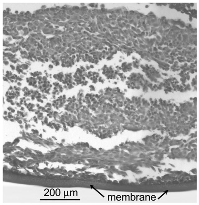
H&E section of multicellular layer grown from the OCM-1 human choroidal melanoma cell line after six days of growth. The membrane is located at the bottom. The thickness of this MCL was 823 ± 20 μm (mean ± SD, 10 measurements).
Official websites use .gov
A
.gov website belongs to an official
government organization in the United States.
Secure .gov websites use HTTPS
A lock (
) or https:// means you've safely
connected to the .gov website. Share sensitive
information only on official, secure websites.
H&E section of multicellular layer grown from the OCM-1 human choroidal melanoma cell line after six days of growth. The membrane is located at the bottom. The thickness of this MCL was 823 ± 20 μm (mean ± SD, 10 measurements).