
Figure 1. IHF-and AHU-DNA cocrystal structures.
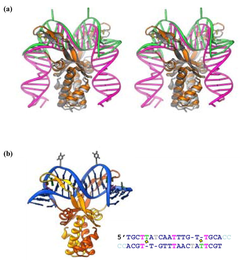
(a) Stereoview of a superposition of the IHF- and AHU-DNA complexes. IHF protein is shown in grey and white while the IHF DNA is pink (1IHF). AHU is gold and the bound DNA is green (1P71). Prolines at the tips of arm-like β -ribbon extension are in yellow. (b) AHU-DNA complex. The AHU-DNA complex (1P71) is color coded as in Table 1. The protein subunits are gold and orange and the intercalating prolines are in yellow. Canonical DNA is blue while unpaired bases are green (stacked) or grey (flipped) and mismatches are pink. Part b reprinted from Figure 3 of Swinger and Rice, 200310.