
Figure 5. EMSA experiment shows that IHF binds to phased single-base insertions.
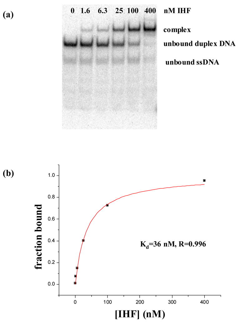
Panel (a) shows a polyacrylamide native gel shift experiment between duplex 4 containing 2 single-T insertions and IHF. Assays were performed in binding buffer, 20mM Tris–HCl (pH 8.0), 70mM NaCl, 1 μ g/ml of salmon sperm DNA, and 5% glycerol, at 4°C by incubation of the 32P-labeled DNA various concentrations of protein. Further details are described in Materials and methods. Panel (b) is the curve fit used to extract an apparent binding constant of 36nM for the gel in panel (a). After three repetitions of the experiment, an apparent Kd of 42±6nM was determined as reflected in Table 1.