
Figure 1.
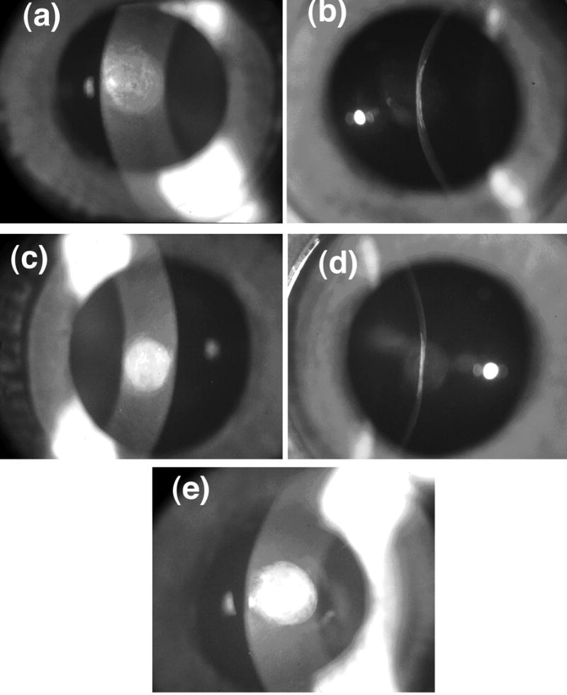
Slit-lamp photographs of scars show that there are substantial variations in scattering intensity across the wound with some regions appearing less opaque than others. The photographs with narrow slit illumination show that the scarred region is flat and thinner than the unwounded cornea. There are variations of scattering intensity with depth, with some regions appearing relatively transparent. (a) Cornea C8L from Group 1 (see Table 1 and Figure 2). (b) Cornea C8R from Group 2. (c) Cornea C25L – the single cornea in Group 3.