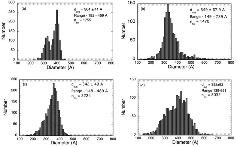
Figure 5.

Fibril diameter distributions from the micrographs in Figures 4 a–d. The distribution from cornea C8LM15983 in (a) is bimodal with distinct peaks at 320A and 390A. Bimodal diameter distributions have not been reported previously for such scars.
