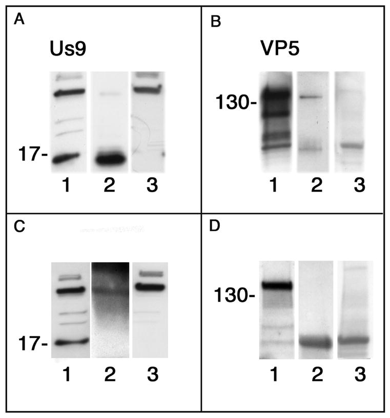
Figure 10.

Comparison of co-immunoassays of lysates of optic pathways of mice infected with wt virus and collected 3 dpi. In each panel lane 1 is a Western blot of lysate before incubation with the beads (positive control), lane 2 is the blot of the elutate fraction and lane 3 is a blot of the eluate fraction from uninfected mouse brain (negative control). A, lane 2 and B, lane 2. We detected both Us9 and VP5 in the lysate affinity assayed with us9 antibody coupled beads. C, lane 2 and D, lane 2. No significant Us9 or VP5 was detected in the lysate affinity assayed with non-specific IgG coupled beads. The molecular weight markers at ~17 kDa indicate one of the multiple forms of Us9. B and D, marker at ~130 kDa.
