
Figure 2.
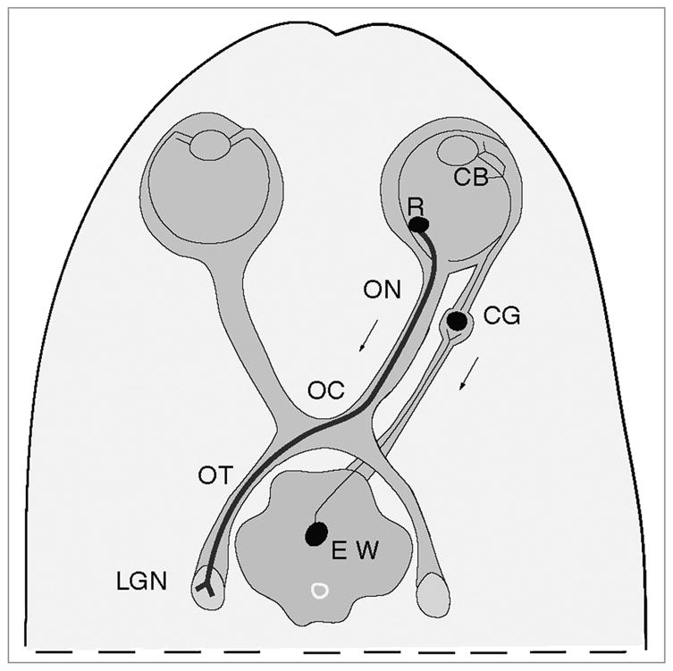
Diagram of the mouse model of infection. By injecting HSV into the vitreal chamber of the eye, we can infect retinal ganglion cell bodies (R). The anterograde transport of virus can be assayed from retinal ganglion cell bodies, along the axons of the cells in the optic nerve (ON), optic chiasm (OC) and optic tract (OT) to the site of termination in the lateral geniculate nucleus (LGN) of the thalamus. As a result of the same injection we can assay the retrograde transport of virus by axons of ciliary ganglion neurons (CG). From the ciliary body in the eye (CB) the virus is transported retrograde by axons of ciliary ganglion neurons (CG) and then transynaptically to the axons of the neurons of the Edinger-Westphal nucleus (EW) in the midbrain.