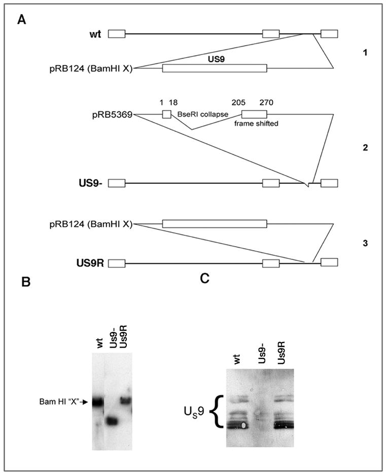
Figure 3.

A. Construction of Us9 mutant viral stains. 1. Cartoon of genome of F strain virus. Us9 is located in the unique short regions. The Bam HI digestion fragment (Bam HI X) was used to produce pRB124. 2. The pRB124 was digested with BseR1 and religated to collapse the region between the two BseRI sites. PRB 5369 was generated. This contained the first 18 nucleotides of the US9 sequence and nucleotides 205–270 were frame shifted. pRB 5369 was used to isolate the mutated virus called US9-. 3. The rescued version was generated by co-infection of cells with US9- DNA and the wt fragment in pRB124. The selected isolate was named Us9R. B. Southern blots of DNA from the viral stocks. The size of fragments from the wt and Us9R are indistinguishable. The Us9- fragment is smaller than the other two strains. C. Western blots of Us9 protein expressed in retinas by the three strains. The immunobands indicating the presence of the several forms of Us9 protein (bracket) are indistinguishable in retinas infected with the wt and Us9R. The Us9 proteins were not expressed in the retinas infected with the Us9- strain.
