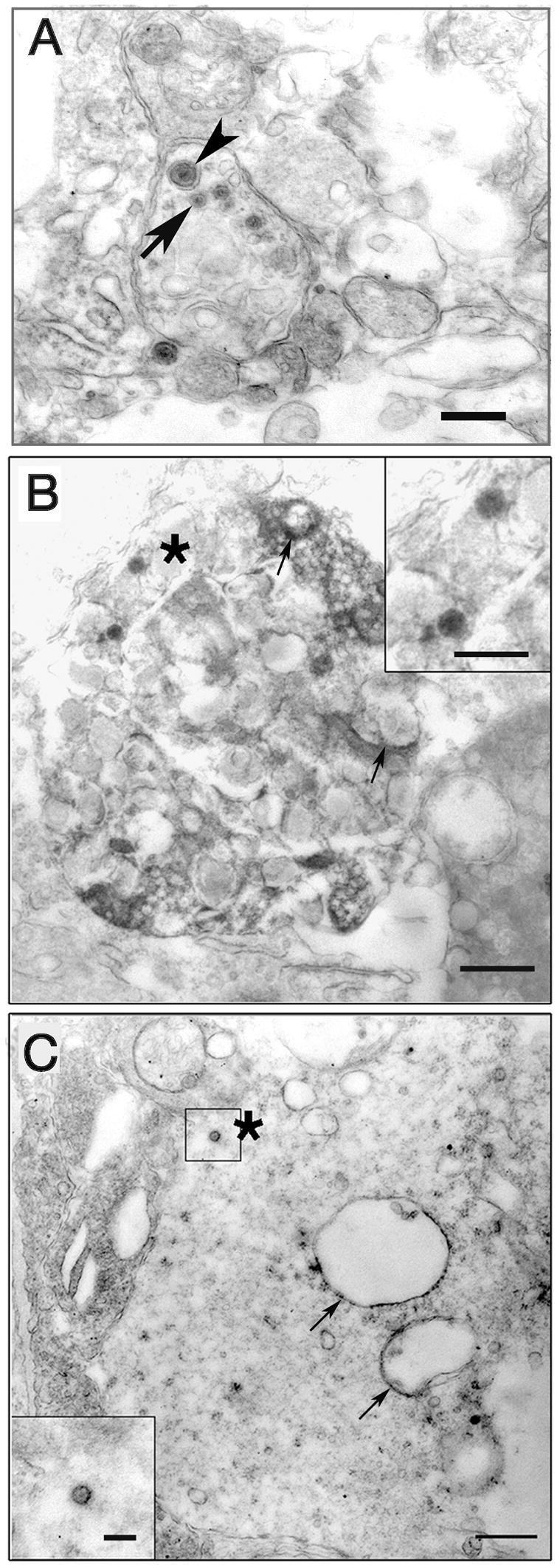
Figure 4.

EM immunohistochemistry of virally infected retinal ganglion cells 24 hr pi. A. Infection with wt strain. Retinal ganglion cell axons are concentrated in a zone immediately adjacent to retinal ganglion cell bodies. Both enveloped (arrowhead) and unenveloped capsids (arrow) can be seen in the axon. Bar = 0.5 μm. B. Retinal ganglion cell infected with Us9R virus. Viral antigen is concentrated on the surfaces of larger vesicles (arrows) as well as on capsids (asterisk) in the cytoplasm. Bar = 0.5 μm Inset, enlarged capsid. Bar = 200 nm. C. Us9- infected ganglion cell body with immunostained vesicles (arrows) and a capsid (asterisk) in the cytoplasm. Bar = 0.5 μm, inset Bar = 200 nm.
