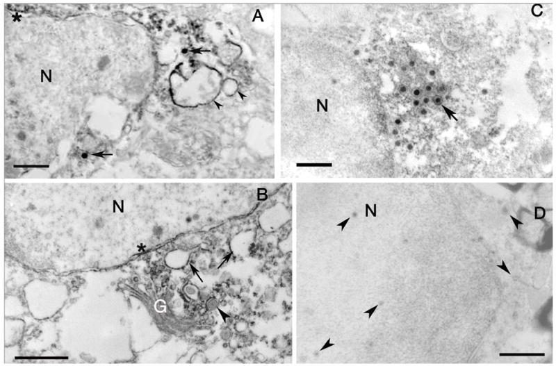
Figure 9.

A. EM immunohistochemistry of Us9 labeling of retinal ganglion cell bodies. The capsids (arrows) in the cytoplasm are immunopositive. The label is also seen over vesicles (arrowheads) and portions of the outer nuclear envelope membrane (asterisk). Bar = 550 nm. B. Portions of the Golgi complex (G) are immunoreactive, as are vesicles (arrows) in the cytoplasm and portions of the nuclear envelope (asterisk). Note that the capsid surrounded by membrane is not immunoreactive (arrowhead). Bar = 1.0 μm. C. EM immunohistochemistry of Us9 labeling of an astrocyte in the optic nerve. Both enveloped and unenveloped immunoreactive capsids (arrow) are concentrated near and within other vesicular structures. The nuclear envelope is also immunolabeled (asterisk). Bar = 550 nm. D. Substituting preimmune serum for the Us9 antibody demonstrates the absence of immunoreactivity and unlabeled capsids (arrowheads). N, nucleus. Bars = 1.0 μm.
