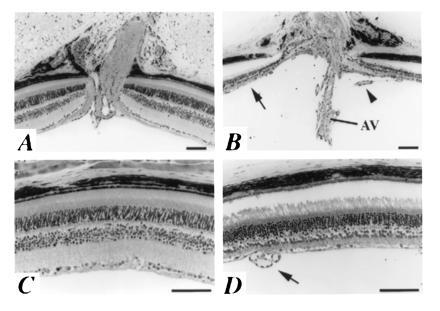
Figure 2.

Eye defects in heterozygous Pax21Neu mice. Retinal morphology of adult (3–6 months of age) wild-type (A and C) and heterozygous (B and D) mice. In comparison to the normal eye (A), note the broad and cup-shaped optic disc in the heterozygote (B) and pronounced thinning of all neural layers of the adjacent retina (arrow). An arterial vessel (AV) projects into the vitreous; a small branch vessel (arrowhead) arches to the surface of the retina. Beyond the immediate region of the optic disc, the inner retinal layers of the heterozygous eye (D) are still markedly thinned, but the photoreceptor layer is similar in thickness to that in the normal eye (C). Preretinal vessels (arrow) are visible all along the inner surface of the mutant retina (D). (Bars = 100 μm.)
