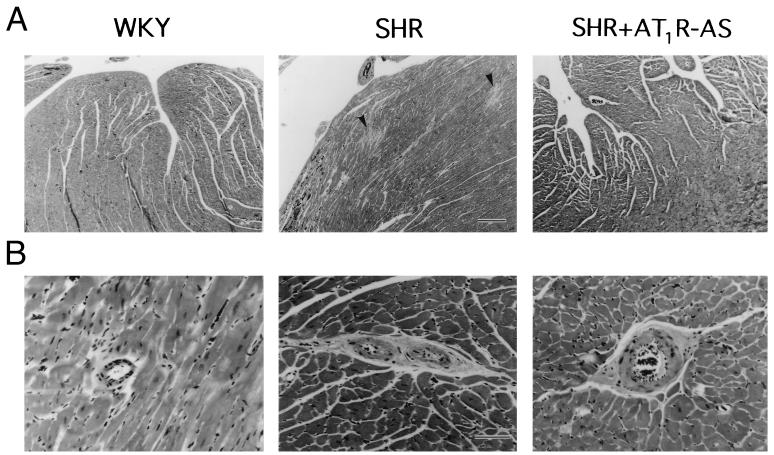
Figure 5.
Effect of AT1R-AS on cardiac and perivascular fibrosis. (A) Photomicrographs of sections taken from left ventricular subendomyocardium from WKY, SHR, and SHR + AT1R-AS, respectively. Photographs were ×50 magnification at ×4 objective. Calibration bar is 12 mm = 250 μm. Arrows in SHR panel indicate multiple focal areas of fibrosis. This phenomenon was not seen in hearts from WKY or SHR + AT1R-AS. (B) Sections from midmyocardium showing small arteries and arterioles (<100 μm diameter) from WKY, SHR, and SHR + AT1R-AS-treated hearts. Photographs were taken at ×308 magnification at ×40 objective. Calibration bar is 15 mm = 50 μm. Vessels from SHR demonstrated dense perivascular fibrosis. This was not seen in vessels from WKY or AT1R-AS-treated rats.

