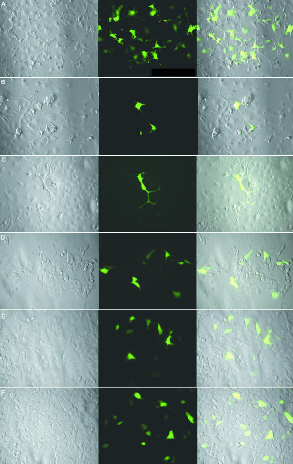
Figure 5.

GFP-XRRA1 fusion protein in COS-7 and HCT116 clones. A, B, and C are transfected COS-7 cells. D, E, and F are transfected HCT116Clone2_XRR cells. Control with GFP cassette alone is on panel A and D. Expression of GFP-truncated XRRA1 is shown in panels B and E. Expression of GFP-full length XRRA1 is shown in panels C and F. Transfected COS-7 cells were examined by fluorescence microscopy after 3 weeks of G418-selection. Expression of GFP-XRRA1 fusion protein was detected only transiently in HCT116 clones. The leftmost panel is a phase-contrast picture of the cells. The middle panel is green fluorescence shown with FITC filter. The rightmost panel is an overlay of the two previous panels. Magnification used was 400×.
