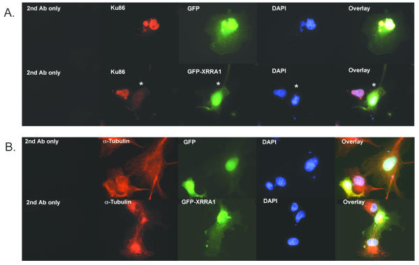
Figure 6.
Immunocytochemistry of COS-7 cells over-expressing GFP-XRRA1 with Ku86 (A) and α-Tubulin (B). (A) Ku86 (red fluorescence) was localized in nuclei. Nuclear Ku86 immunostaining was eliminated (asterisk) in the presence of GFP-XRRA1 (green fluorescence), but not with the GFP alone. (B) Neither GFP nor GFP-XRRA1 changed the immunostaining of α-Tubulin (red fluorescence). DAPI-stained nuclei are shown in blue fluorescence. Magnification used was 1000×.

