
Figure 2.
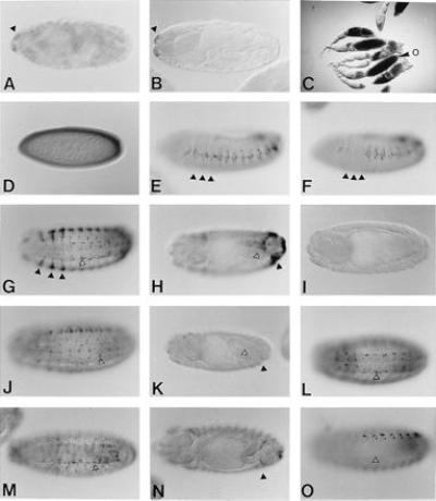
Enhancer activity of the Hoxa-7 intron in Drosophila. Stainings of embryos with antiserum against β-gal, if not indicated differently. (A) Stage 17 embryo of p40 showing epidermal expression in the maxillary segment (arrowhead). (B) Embryo of a control Hz50pl line without insert shows the same expression as A. (C) β-Gal activity staining of PB6 ovaries. Strong staining is obtained in the nurse cells from at stage 9 on and later in the oocyte (o). (D) Stage 5 PB6 embryo showing maternal expression. (E–N) Stage 13–15 embryos (E) PS4 embryo showing expression in the lateral epidermis. The thoracic segments are indicated by arrowheads. (F) PS4 embryo in a presumptive Antpw10 mutant background. Expression in the lateral epidermis in the three thoracic segments is missing (arrowheads). (G and H) XS3 embryos showing ventrolateral expression (G) and expression in the posterior segments (H). The zygotic staining in PS4, XS3, and FS2 lines is identical to PB6. (I) HB1 embryos show no expression. (J and K) XO3 embryos showing staining in cells of the ventral and peripheral nervous system and in some lateral epidermal cells (arrowhead) (J), but not in the cells around the posterior midgut (open arrowhead) and in the posterior segments (solid arrowhead) (K). (L) P1 embryo showing staining in the ventral nervous system. (M and N) ON3 embryo showing the same staining as P1 (M) as well as weak staining in the posterior segments (N). (O) HA1 embryo showing similar (nuclear) staining as HS1, but the P1 pattern (L) in the ventral nervous system is absent (arrowhead).