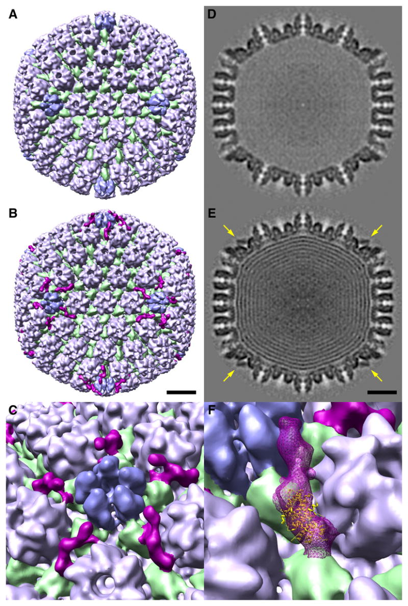
Figure 2. The CCSC is present on C-capsids but not on UL25-null A-capsids.

Surface renderings of cryo-EM reconstructions are shown in B (C-capsid) and A (UL25-null A-capsid). Triplexes are green; hexons (UL19 hexamers with six VP26 molecules around their outer tip) are light blue; pentons (UL19 pentamers) are mid-blue. The CCSC in B is purple. The capsids are viewed along a 2-fold axis of icosahedral symmetry. C shows a blow-up of the region around a penton, presenting five different views of the CCSC molecules clustered around this vertex. Bar (B, E) = 20 nm. D and E show central sections of the two capsids. High density is dark. The UL25-null A-capsid is empty; the C-capsid contains nested shells of DNA (Booy et al., 1991). Arrows in E point to the CCSC which is sampled in this plane. F shows the optimal fit of a UL25 fragment into the CCSC.
