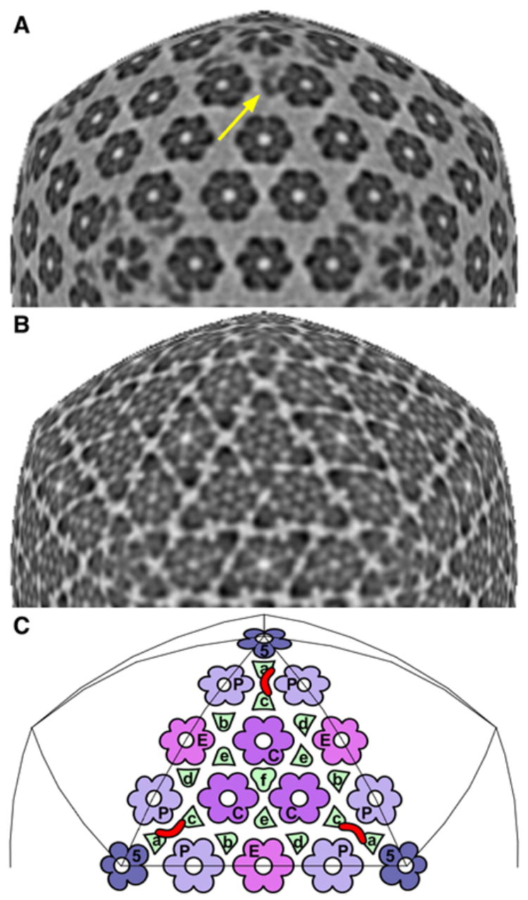
Figure 3. The CCSC is visible at only one of six quasi-equivalent sites on the HSV-1 C-capsid.

A & B show icosahedral sections through the C-capsid at radii of 61 nm and 53 nm, respectively, along the 3-fold axis. In them, the sampling surface maintains the same radial position relative to the capsid shell (as opposed to the capsid center). Section A is rescaled so that features in it line up with underlying features in section B. Section A samples the CCSC (yellow arrow), whereas section B samples the triplexes as well as the capsomer protrusions, just above the floor. In C are mapped the positions of the four kinds of capsomers and six kinds of triplexes on one triangular facet. Each CCSC (one is marked in red) overlies a pair of triplexes (Tc, Ta). Other quasi-equivalent sites may be found by substituting another kind of triplex for Tc. There are 320 triplexes, but maximum CCSC binding would correspond to 160 copies, because one CCSC occludes two triplexes. The CCSC is seen on C-capsids at only one of the six possible sites, at 50% occupancy. This occupancy may increase in virions, whose UL25 content is higher than that of C-capsids (Thurlow et al., 2006). Bar (B) = 15 nm.
