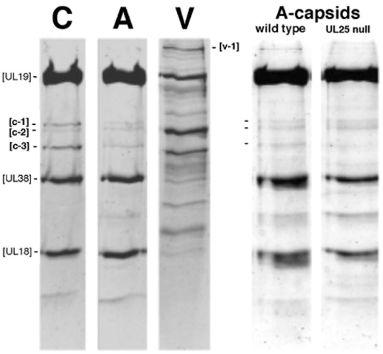
Figure 5. Identification of minor protein components of HSV-1 capsids.

At left, C-capsids (C), wildtype A-capsids (A), and virions (V) are compared by SDS-PAGE with Coomassie Blue staining. Equal amount of protein were loaded in each lane. The major capsid proteins, UL19, UL38 and UL18 contribute strongly staining bands. The weak bands, c1, c-2, and c-3 were identified by mass spectrometry as UL17, UL6, and UL25, respectively. The v-1 band was identified as UL36. At right, wild-type A-capsids are compared with A-capsids from a UL25-null mutant. Again, c1, c-2, and c-3 are marked. The mutant capsids do not contain UL25, as confirmed by Western blotting (McNab et al., 1998). The very faint band running just below the UL25 position in the UL25-null mutant lane represents an unknown contaminant.
