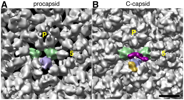
Figure 6. Changes affecting the CCSC binding site on maturation of the HSV-1 procapsid.

The regions of capsid surface surrounding the CCSC binding site are compared in cryo-EM reconstructions at ~ 2 nm resolution of the procapsid (A – from Cheng et al., 2002) and the C-capsid (B). In both cases, the two triplexes and one UL19 subunit that form the binding site for a single representative CCSC are shaded green and blue, respectively; this CCSC is purple in panel B and the UL35 subunit that tops the interacting UL19 subunit (but is not present on the procapsid) is gold. As reference points, the 5-fold axis of the adjacent UL19 penton is marked “5”, and a neighboring P-hexon with P. Bar = 10 nm.
