
Figure 6.
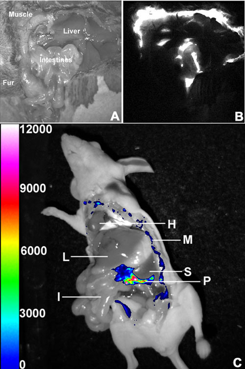
CMV::luc expressed in pancreas and only a few other areas. (A) Reference image of dissected adult CMV::luc mouse with four areas labeled. (B) Expression from the same mouse. (C) Dissection of Hr-CMV mouse. Shown is an overlay of the bioluminescence image on the reference image to identify brightest tissues. Pancreas (P) revealed high expression and muscle tissue (M) showed weak signal, but heart (H), liver (L), spleen (S), and intestine (I) did not.