
Figure 2.
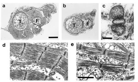
Morphology of E18 β1-null fetuses shows a reduction in muscle mass and disorganization of the myofibrils. (a and b) Transverse sections through the hindlimbs of a control (a) and β1-null (b) fetus. (Bar = 0.3 mm.) Sections were cut at approximately the same position and the tibia (T) and fibula (F) can be seen. The amount of muscle in the β1-null fetus is markedly decreased. (c) Electron micrograph of a triad from a β1-null fetus. (Bar = 0.1 μm.) (d and e) Electron micrograph of muscle from control (d) and β1-null (e) fetus. (Bar = 1.2 μm.)