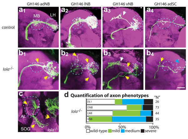
Figure 3.
Axon wiring defects in lola-/- MARCM clones. Representative confocal images of (a) wild-type control and (b) lola-/- adNB (1), lNB (2) vNB (3) and DL1 SC (4) clones showing PN axons near their normal target: the mushroom body (MB) and lateral horn (LH), both marked by blue dotted circles. (c) Example of an axon mistargeting to SOG after exiting the posterior AL (blue dotted circle). Double-headed white arrow demarks dendritic mistargeting to SOG. Blue arrows denote a lack of correct targeting while yellow arrows demark ectopic branching. anti-CD8::GFP in green, anti-nc82 neuropil in magenta. Scale bar, 20 μm. (d) Quantification of severity of axon targeting defects. Mild: axons target correctly but have ectopic branches (b1, b2); medium: axons target to the correct region but follow an incorrect trajectory or bifurcate (b3, b4); severe: axons target to the SOG or abnormal brain regions (c).

