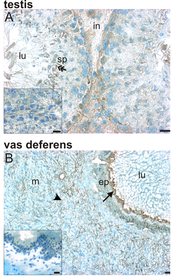
Figure 4.
Localization of Rxfp2 protein in the testis (A) and vas deferens (B). Immunohistochemistry for Rxfp1 (1:100 dilution of primary antibody) was done in cryosections of the testis and vas deferens; sections were counterstained with methylene blue. Inserts show controls incubated with non-immune rabbit serum instead of primary antibody. Scale bars are 5 μm. Results are representative of at least 3 independent determinations, each with a different animal. ep = epithelial layer, in = interstitium, lu = lumen, m = muscular layer, sp = spermatid. Arrows indicate labeled epithelial cells, arrow heads indicate labeled muscular cells, and double-headed arrows indicate labeled spermatids.

