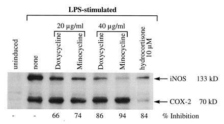
Figure 4.

Western blot analysis of iNOS in RAW 264.7 cells exposed to doxycycline or minocycline in the presence of LPS. RAW 264.7 cells activated with LPS (100 ng/ml) for 16–18 h, with and without doxycycline or minocycline (20–40 μg/ml) and hydrocortisone (10 μM) were analyzed for total iNOS and COX-2 protein. Briefly, 30 μg of cell extract was loaded onto SDS/PAGE gels and the filter probed simultaneously with anti-iNOS and anti-COX-2 mouse mAb followed with an anti-mouse serum conjugated to horseradish peroxidase on the same filters. Bands were quantitated on a densitometer and the percent inhibition was compared with the LPS-stimulated cells. Data represent one of four similar experiments.
