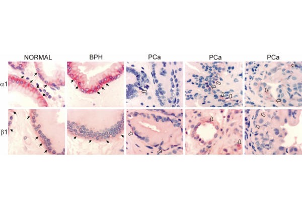
Figure 2.
Immunohistochemical staining of Na, K-ATPase α1 and β1 subunits in canine normal, BPH and PCa tissues. Sites of Na, K-ATPase immunoreactivity are stained red (using Fast-Red TR/Naphthol AS-MX as precipitating substrate for the alkaline phosphatase conjugated secondary antibody). In normal and BPH sections Na, K-ATPase immunostaining is clearly visible in the basolateral membrane domain of epithelial cells (arrows). In three different PCa specimens Na, K-ATPase expression levels appear to be significantly lower than in normal or BPH tissues and Na, K-ATPase is diffusely spread within focal areas of the neoplasm. Nuclei were counterstained with haematoxylin.

