Figure 3.
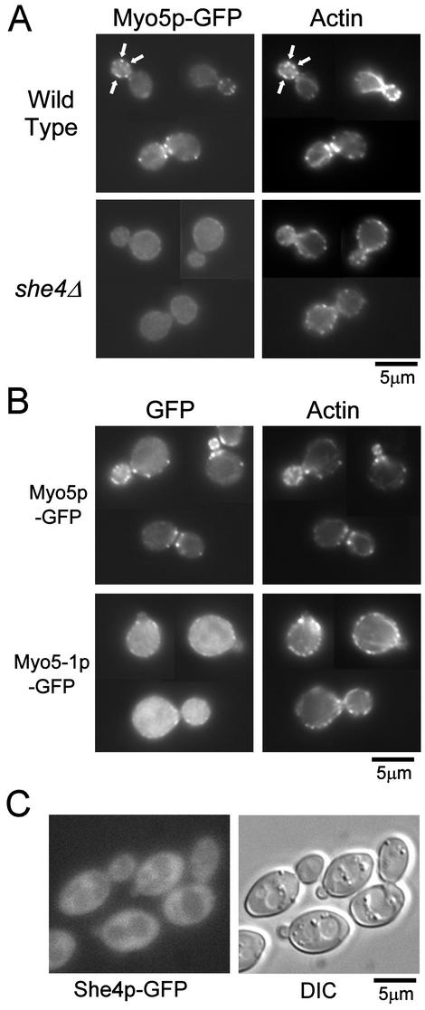
She4p is required for proper localization of Myo5p-GFP and F-actin. (A) Localization of Myo5p-GFP in she4Δ cells. Myo5p-GFP expressing cells, YKT159 (wild-type) and YKT288 (she4Δ) were grown at 25°C, fixed and stained with TRITC-phalloidin. Localization of Myo5p and F-actin was visualized with a GFP band pass filter (Myo5p-GFP) and a TRITC filter (actin). For each observation, the same exposure and processing parameters were used. Arrows indicate examples of colocalization of Myo5p-GFP and actin patches. (B) Localization of Myo5-1p-GFP. YKT114 cells were transformed with plasmids, pKT1235 (Myo5p-GFP) or pKT1403 (Myo5-1p-GFP). Cells were grown in SD-Leu liquid medium at 25°C, incubated at 37°C for 1 h, fixed, stained, and observed as described in A. (C) Localization of She4p-GFP. She4p-GFP–expressing cells, YKT274, were grown at 25°C, and She4p-GFP was visualized using a GFP band pass filter (She4p-GFP) or differential interference contrast optics (DIC). Bars, 5 μm.

