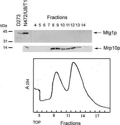
Figure 4.
Analysis of yeast mitochondrial ribosomes. Mitochondria of the wild-type strain W303-1A were lysed with 1% potassium deoxycholate and clarified by centrifugation at 14,000 × gav for 15 min. Mitochondrial ribosomes were enriched by centrifugation of the extract through a 50% sucrose cushion (Myers et al., 1987). The ribosomal pellet was suspended in AMT (500 mM ammonium chloride, 10 mM MgCl2, 10 mM Tris-Cl, pH 7.5, 6 mM β-mercaptoethanol) and was layered on a 5-ml column of a 10–30% linear sucrose gradient in AMT buffer. After centrifugation at 65,000 rpm in a Beckman SW65Ti rotor for 100 min, the gradient was fractionated into 18 fractions. Only fractions containing the large and small subunits were analyzed as in Figure 3 with antibody against Mtg1p and Mrp10p (Jin et al., 1997).

