
Fig. 9. Activation of ERK1/2 during cisplatin exposure in RPTC.
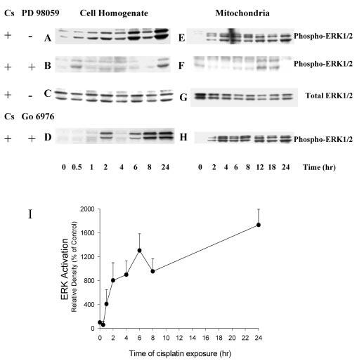
RPTC were treated with 50 μm cisplatin, and samples were taken at 0, 0.5, 1, 2, 4, 6, 8, 12, 18, and 24 h for measurements of protein levels of phosphorylated and total ERK1/2 using immunoblotting. Immunoblotting was performed as described in the legend to Fig. 8. A–D, protein levels of phospho- and total ERK1/2 in RPTC homogenates. E–H, protein levels of phospho- and total ERK1/2 in RPTC mitochondria. PD98059 (50 μm) and Go6976 (10 nm) were added 1 h prior to cisplatin treatment. Presented data are representative of three independent experiments (cell isolations). I, cisplatin-induced ERK1/2 activation quantified by densitometry. Results are the average ± S.E. of three independent experiments (RPTC isolations).