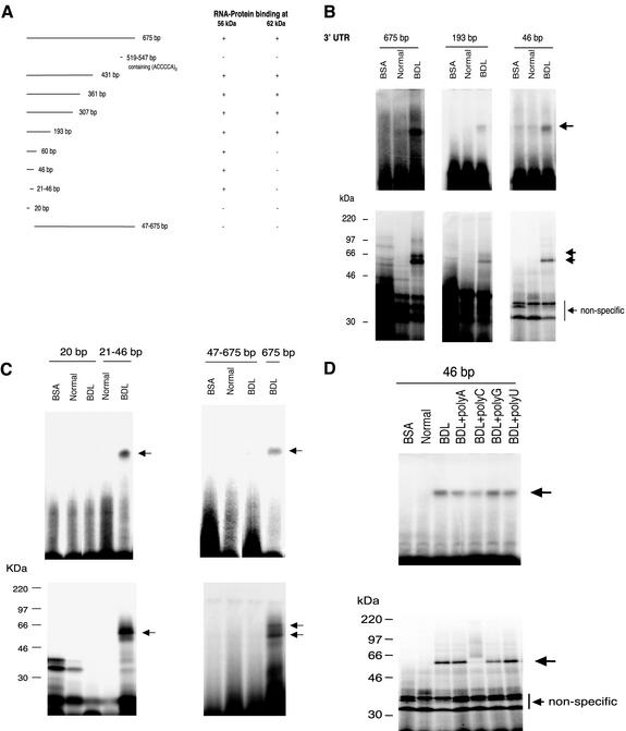
Figure 3.
RNA binding proteins bind different ECE-1 3′ UTR sequences. (A) Multiple 3′ UTR fragments were cloned as described in MATERIALS AND METHODS. Protein binding activity was determined by gel mobility assay and UV cross-linking as in MATERIALS AND METHODS and is reported diagrammatically. (B and C) Gel mobility assay (top panels) and UV cross-linking experiments (bottom panels) examining specific areas of the UTR are shown. Cellular cytoplasmic extracts were incubated with the indicated 32P-labeled fragments as described in MATERIALS AND METHODS. Arrows indicate RNA–protein complexes. Bovine serum albumin was used as a negative control. The figures are each representative of four different experiments. (D) Cellular cytoplasmic extracts were incubated with a 32P-labeled 46-base pair fragment of ECE-1 3′ UTR as described in MATERIALS AND METHODS. Poly(A), (C), (G), or (U) (200 ng) was added and RNA–protein complexes were analyzed by gel mobility assay (top). The lower panel shows UV cross-linking. Arrows indicate RNA–protein complex. Bovine serum albumin was used as a negative control. The figure is representative of six different experiments.

