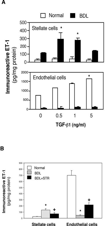
Figure 6.
Effect of TGF-β1 on ET-1 production in stellate and endothelial cells in vitro and in vivo. (A) Stellate and sinusoidal endothelial cells were isolated from normal rat livers or those injured by BDL as in MATERIALS AND METHODS. After adherence for 48 h, serum-free medium along with TGF-β1 was applied to isolated cells at the indicated concentrations was introduced for 24 h. ET-1 in conditioned supernatants was measured by radioimmunoassay as in MATERIALS AND METHODS and normalized to the total protein content in the monolayer. *p < 0.05 compared with medium without TGF-β1 (n = 5). Note that for stellate cells, ET-1 production by cells after BDL was significantly greater than in normal cells at all TGF-β concentrations. (B) Stellate and endothelial cells were isolated from normal rat livers, those after BDL, or after exposure to an STR during the induction of liver injury by BDL as in MATERIALS AND METHODS. After isolation, cells were allowed to adhere for 48 h, serum-free conditions were introduced for 24 h, and ET-1 in conditioned medium was measured by radioimmunoassay. *p < 0.05 compared with normal and +p < 0.05 compared with BDL (n = 8).

