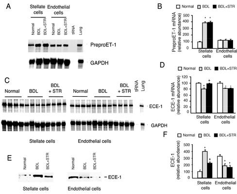
Figure 7.
Effect of TGF-β1 on precursor ET-1 and ECE-1 in vivo during hepatic wound healing. Stellate and endothelial cells were isolated from three groups of rats as described in Figure 6. (A) Total cellular RNA was isolated and preproET-1 mRNA was detected by RNase protection assay as in MATERIALS AND METHODS. (B) Specific preproET-1 bands were normalized to the signal for GAPDH; the level of preproET-1 mRNA from normal stellate cells was set at 100. (C) Cellular ECE-1 mRNA was detected by RNase protection assay. (D) Specific ECE-1 bands were normalized to the signal for GAPDH, and ECE-1 mRNA from normal stellate cells was arbitrarily set at 100. (E) ECE-1 from the same cells was detected by immunoblot as in MATERIALS AND METHODS. (F) Specific bands were quantitated, the level of ECE-1 from normal stellate cells was arbitrarily set at 100, and data are presented graphically. *p < 0.05 compared with normal and +p < 0.05 compared with BDL (n = 4).

