
Figure 2.
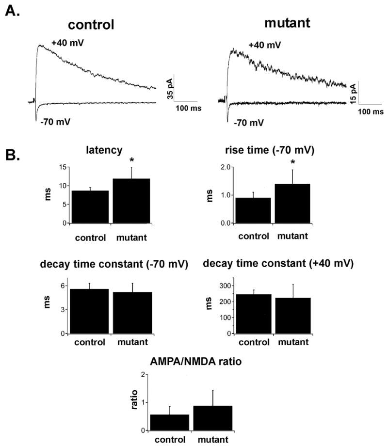
A. Thalamocortical EPSCs evoked at −70 and +40 mV in slices obtained from control (left) and mutant (right) animals at P4–P5. Stimulation artifacts have been blanked. Note that both slowly decaying NMDA-receptor mediated currents and rapidly decaying non-NMDA mediated currents can be evoked in slices obtained from both control and mutant animals. B. Bar graphs of latency, rise time, decay time constant (at −70 and +40 mV), and AMPA/NMDA ratio of thalamocortical EPSCs evoked in slices obtained from control or Emx1-cre;Dnmt1 mutant animals at P4–P6. Asterisks denote statistical significance at p<0.05 level. Note that thalamocortical EPSCs in mutants have slightly more prolonged latencies and rise times, but the decay time constant at −70 and +40 mV, as well and AMPA/NMDA ratio is not statistically different between the two groups.